What Does Breast Cancer Look Like On An X Ray / Dual-energy beats bone suppression in chest imaging : It can also be used to investigate the cause of breast problems, such as a breast mass, pain and nipple.
Dapatkan link
Facebook
X
Pinterest
Email
Aplikasi Lainnya
What Does Breast Cancer Look Like On An X Ray / Dual-energy beats bone suppression in chest imaging : It can also be used to investigate the cause of breast problems, such as a breast mass, pain and nipple.. The breast cancer can either be: Microcalcifications look like tiny white specks, usually clustered together. A sudden, noticeable new pain is the most common symptom of cancer that has spread to the bone. It looks for early changes that could be a sign of cancer. The milk ducts carry your breast milk from lobules, where milk is produced, to your nipple.
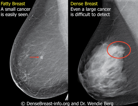
It is also an anatomic test, which means cancer is detected based on changes in how the breast anatomy looks. Cancer cells can remain within the milk ducts and this is considered as noninvasive cancer or ductal carcinoma in situ. 1 the gray areas correspond to normal fatty tissue, while the white areas are normal breast tissue with ducts and lobes. Paget's disease is usually a sign of breast cancer in tissue behind the nipple, or breast tissue away from the nipple. The breast cancer can either be:
Undiagnosed primary lung carcinoma with initial ... from www.cancerjournal.net Although breast cancer can spread to any bone, the most common sites are the ribs, spine, pelvis, and long bones in the arms and legs. Your skin may also become very sensitive and itchy. The radiographer positions one breast at a time between two flat plates on the machine. That indicates that the lesion likely contains a variety of elements, which may or may not indicate breast cancer. When breast cancer moves into the lung, it often doesn't cause symptoms. The american cancer society says. A tumor that is benign, it is not a health problem and it may not grow or change shape. A lump or tumor will show up as a focused white area on a mammogram.
It can also be used to investigate the cause of breast problems, such as a breast mass, pain and nipple.
Spaying before the first heat cycle will greatly reduce your dog's risk for developing breast cancer. The breast cancer can either be: What does breast cancer look like on a mammogram? 1 the gray areas correspond to normal fatty tissue, while the white areas are normal breast tissue with ducts and lobes. Paget's disease is usually a sign of breast cancer in tissue behind the nipple, or breast tissue away from the nipple. When possible, the doctor reading your mammogram will compare it to your old mammograms. Microcalcifications also are often associated with benign conditions, but their appearance in clusters or lines may indicate the presence of cancer. The breast is compressed between two firm, flat surfaces to spread the tissue out. The earlier you find the disease, the easier it is to treat. Tumors may be benign or cancerous. Early spaying is the best method for prevention of this form of cancer. This compression helps to give a clear picture. Although breast cancer can spread to any bone, the most common sites are the ribs, spine, pelvis, and long bones in the arms and legs.
Doctors use a mammogram to look for early signs of breast cancer. A tumor that is benign, it is not a health problem and it may not grow or change shape. 1 the gray areas correspond to normal fatty tissue, while the white areas are normal breast tissue with ducts and lobes. As it travels, different parts of the body absorb different amounts of the energy. It looks for early changes that could be a sign of cancer.
Breast Density | Keep Healthy from www.keep-healthy.com The lungs are a common site for breast cancer metastases. A sudden, noticeable new pain is the most common symptom of cancer that has spread to the bone. The radiographer positions one breast at a time between two flat plates on the machine. Ct, mri, and pet scans. It looks for early changes that could be a sign of cancer. It is also an anatomic test, which means cancer is detected based on changes in how the breast anatomy looks. The radiographer positions one breast at a time between two flat plates on the machine. A radiologist will look at your mammogram.
Any area that does not look like normal tissue is a possible cause for concern.
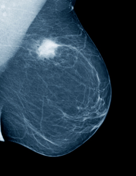
Early spaying is the best method for prevention of this form of cancer. Your skin may turn pink, red, tanned, or look like it has sunburn. Macrocalcifications look like large white dots or lines. This can help show if any findings are new, or if they were already there on previous mammograms. It can detect breast cancer long before a tumor might be felt by you or your provider. A lump or tumor will show up as a focused white area on a mammogram. Abnormalities such as cancerous tumors usually appear brighter because they are denser. A mammogram can show breast changes such as calcifications, masses, or other symptoms that might be cancer. The american cancer society says. It looks for early changes that could be a sign of cancer. The most important screening test to detect breast cancer early is the mammogram. Any area that does not look like normal tissue is a possible cause for concern. That indicates that the lesion likely contains a variety of elements, which may or may not indicate breast cancer.
The radiographer positions one breast at a time between two flat plates on the machine. Dense breast tissue can look light gray or white on a mammogram. The lungs are a common site for breast cancer metastases. Doctors use a mammogram to look for early signs of breast cancer. Microcalcifications look like tiny white specks, usually clustered together.
What is a Mammogram & How Does It Detect Breast Cancer? from www.maurerfoundation.org Your skin may also become very sensitive and itchy. For more than half of women who develop stage iv breast cancer, the bones are the first site of metastasis. Dense breast tissue can look light gray or white on a mammogram. Fat and muscle absorb less, so they show up in different shades of gray. When breast cancer moves into the lung, it often doesn't cause symptoms. Abnormalities such as cancerous tumors usually appear brighter because they are denser. Any area that does not look like normal tissue is a possible cause for concern. 1 the gray areas correspond to normal fatty tissue, while the white areas are normal breast tissue with ducts and lobes.
It is also an anatomic test, which means cancer is detected based on changes in how the breast anatomy looks.
It is also an anatomic test, which means cancer is detected based on changes in how the breast anatomy looks. The most important screening test to detect breast cancer early is the mammogram. The milk ducts carry your breast milk from lobules, where milk is produced, to your nipple. A tumor that is benign, it is not a health problem and it may not grow or change shape. The most common type of breast cancer is ductal carcinoma, which forms in the linings of the milk ducts within the breast. Rapidly growing cells, such as cancer cells, are more susceptible to the effects of radiation therapy than are normal cells. The breast cancer can either be: When breast cancer moves into the lung, it often doesn't cause symptoms. Learn about the causes, symptoms, treatment, and more. The american cancer society says. Ct, mri, and pet scans. The earlier you find the disease, the easier it is to treat. A radiologist will look at your mammogram.
Cancer Council Nsw Address - Cancer Council NSW's Weather & UV Chart on Vimeo / Donate to cancer council and help pave a brighter future for rylee and other cancer patients. . Cancer council nsw goal is to reduce deaths from cancer by 50 over the next 20 years. 4 prince of wales clinical school, unsw, new south wales, australia(1); Donate to cancer council and help pave a brighter future for rylee and other cancer patients. Ms helen gooden, ms marie malica, mr andrew penman, ms monica robotin, a/prof freddy sitas, and ms nysha thomas. There's an exhaustive list of past and present employees! Cancer research division, cancer council nsw, new south wales, australia(2). Through the development of prevention strategies, research into new treatments and cures, and by providing clinical and emotional support to those affected by cancer. Cancer council nsw stands as one of the best qualified companies among its field. View all our cancer council nsw vacancies n...
Signs And Symptoms Of Urinary Bladder Cancer - Diagnosing Bladder Cancer 1 - Signs, Symptoms & Tests ... : Oftentimes, though, these are merely symptoms of a urinary tract infection and antibiotics become the first line of treatment. . The detrusor muscle is the thick muscle deep in the bladder wall. For most people, the first symptom of bladder cancer is blood in the urine, also called hematuria. Blood in the urine (hematuria) is often the most common sign of bladder cancer. There are other symptoms to watch for as well. Bladder cancer is one of the most common types of cancer. For example, blood in the urine is most often caused by a bladder or urinary tract infection or a kidney stone. Patients with urinary incontinence suffer urine leakages or dribbling of urine in the most unexpected moments or a sudden urge to urinate that is impossible to hold back. This is the most common symptom of bladder cancer and occurs in the vast majority of people with bladder can...
Ideas For Making Teachers Day Card - Teachers Day Card Instructables - Actually i am writing on this day so. . You surely would have come across a teacher who understand you always and is great when how to make it nothing can be easier than making this card. Teacher who is taking information and network security. In this video, i am going to show you special cards making at home.please like the video, if you liked the card. Whether it is teacher's day, national teacher day, or teacher appreciation week, make, print, or customize your very own handmade card for that special it was made for teachers day but it can surely be used for other occasions too. See more ideas about teachers day card, cards handmade, inspirational cards. #handmadecard #card #popupcard subscribe to my channel & enjoy. How to make teacher's day card teacher's day greeting pop up card tutorial. Many students put their efforts into making a teachers' day card unique and commi...


Komentar
Posting Komentar